

Bristol Knee Clinic

| + home | |
| + news | |
| + research | |
| + patient information | |
| + the clinic | |
| + the surgeon | |
| + sport physiotherapy | |
| + sports advice | |
| + medico legal | |
| + products | |
| + resources | |
| + contact | |
| + maps | |
| + directions | |
| + site map |
The Bristol Knee Clinic |
Arthroscopy Of The Shoulder - Surgery
Arthroscopic Surgery of the Shoulder
Diagnostic arthroscopy is a well recognized procedure providing orthopaedic surgeons with an exact diagnosis which is not possible with examination and X-ray alone. Patients with non-specific signs, such as partial rotator cuff repairs, are difficult to diagnose and many shoulder conditions can be treated with the development of arthroscopic surgical repair. These methods of treatment continue to develop, offering clear advantages compared to open procedures, such as being less intrusive and offering early recovery for the patient.
Although the development of techniques for shoulder arthroscopy succeeded knee procedures by several years, by now, diagnostic arthroscopy in the shoulder joint is well established. The primary value of arthroscopy for management of the injured shoulder has been in enabling the orthopedists to establish a definite diagnosis, often elusive in this complex joint despite thorough physical examination, radiography, and arthrography. Partial rotator cuff tears typify these difficult-to-diagnose disorders that present with unspecific findings. In recent years, the utility of the arthroscope in shoulder disorders has been extended beyond enhanced diagnostic capability by the development of surgical techniques to treat a wide variety of such disorders, including impingement syndrome. These techniques are evolving as part of a learning curve, similar to that experienced in the development of arthroscopic knee surgery.
The technique we will describe for arthroscopic subacromial decompression is the equivalent of standard methods of anterior acromioplasty performed as open surgery. Although technically demanding, the arthroscopic technique offers distinct advantages, providing prompt visualization of rotator cuff injuries and a less invasive procedure, with maintenance of the deltoid muscle and consequently early rehabilitation.
Patient Selection
Subacromial decompression - Patients with rotator cuff tendonitis or an enlarged overhanging or damaged arcromium bone are ideal for subacromial decompression when conservative measures such as anti- inflammatory drugs and exercise has been unsuccessful in treating the symptoms.
Patients with a complete tear of the rotator cuff may also benefit from this procedure although reconstruction or direct repair of the defect or tear in the rotator cuff is often also required. Patients of middle age between 40 and 60 years with small rotator cuff tears and no weakness may also be appropriate for a simple subacromial decompression procedure rather than additional direct repair of the rotator cuff. The advisability of surgical repair in these patients is a decision for the surgeon in consultation with patients with the results of an MRI scan.

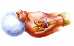
The position of the patient for shoulder arthroscopy and the access points or portals used.
Technique
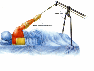
General anaesthetic is used and the patient placed in the lateral position on the operating table tilted 30° posteriorly. A bean bag is used to stabilize the body and the arm is suspended in 30° of abduction and 15° of forward flexion, with 10 pounds of traction applied, Alternately a beach chair position can be used with the patient lying on their back. The acromion, scapular spine, clavicle, acromioclavicular joint, coracoid, and humeral head are marked. Portals are created by making small skin incisions at these sites, and then inserting first a sharp, and then a blunt obturator. Figure 2 shows the portals used for intra-articular and subacromial surgery.

The patient position for shoulder arthroscopy with traction applied to the arm.
Shoulder Intra-Articular Examination and Surgery
First, the posterior portal is formed, and then the anterior portal created using the Wissinger Rod technique.
The joint is examined at the intra-articular portion and the status of articular cartilage on the glenoid and humeral head noted. A electronic shaver can be used to debride and remove any synovitis, Minimal debridement is suggested if the biceps tendon is frayed, in order to preserve this tendon, which functions as a depressor of the humeral head.
The undersurface of the rotator cuff is evaluated and frayed edges debrided. Slight scuffing on the cuff or large fragments that extend into the substance of the supraspinatus tendon can be associated with more extensive disease. This area is debrided of loose tissue, but complete excision of necrotic tissue is not undertaken.
Subacromial Examination and Surgery
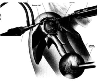
Anterior acromioplasty and decompression of the subacromial space is the aim of the procedure. Decompression is achieved by resecting the subacromial bursa and sectioning the coraco-acromial ligament acromial attachment. The anterior acromioplasty removes the underside of the bone of the acromial arch to provide more room for the rotator cuff tendon. At the end of the procedure the wounds are usually sutured or closed with adhesive strips and dressings applied. It is not uncommon for the shoulder to be swollen from local absorption of fluid into the tissues around the shoulder joint. This usually settles over a few hours as the fluids reabsorb. A sling is usually applied to rest and support the shoulder.

Fig. 2. The position of the arthroscope and electronic shaver for busal arthroscopy and subacromial decompression.
< BACK to Non-operative Treatment | NEXT: Recovery and Rehabilitation >
Related Links..
+ How to make an appointment
+ Arthroscopy Of The Shoulder - see all links
+ Patient Information Home
+ See the clinic
+ More about Mr Johnson
+ top
© The Bristol Orthopaedics and Sports Injuries Clinic 2003. The Bristol Knee Clinic is a trading name of the Bristol Orthopaedic Clinic Ltd. privacy / copyright | contact | Powered By Create Medical


