


Bristol Knee Clinic
| + home | |
| + news | |
| + research | |
| + patient information | |
| + the clinic | |
| + the surgeon | |
| + sport physiotherapy | |
| + sports advice | |
| + medico legal | |
| + products | |
| + resources | |
| + contact | |
| + maps | |
| + directions | |
| + site map |
The Bristol Knee Clinic |


Early Arthritis Of The Knee - Surgical Treatments
Common Surgical Procedures
Arthroscopy
Arthroscopy is the technique of performing surgery inside the knee joint through a telescope without disrupting the surrounding structures. Looking inside the joint with the arthroscope allows the Surgeon to look directly at the menisci, cartilages, ligaments and the articular surfaces. In addition surgery can be undertaken through the arthroscope to remove the damaged area of the joint.
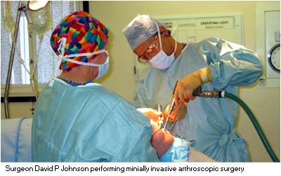
Arthroscopy is commonly performed as a day case procedure with a rapid recovery and a rapid return of function often within a few days. The procedure is very safe and the most common surgical procedure now performed. Where the symptoms originate from a torn cartilage the procedure may be a success in as much as 98% of patients.
Arthroscopic Debridement Of The Knee
An arthroscopic "clean up" or debridement of the damaged surfaces of the knee can in the majority of cases diminish the level of pain and may slow down the rate of progression of the arthritis. Partial removal of the worn cartilage is often necessary and undertaken at the same time, the rough edges of the articular surface are smoothed out and any loose pieces in the joint can be removed. This often has an excellent result with loss of the pain and a return to the previous level of activity. However, because of the underlying problem of wear the symptoms may return in the future, albeit not perhaps for many years. The duration of the improvement in the symptoms is entirely dependent upon the extent and stage of the underlying arthritis. If the symptoms return the arthroscopic debridement may be repeated at intervals but eventually more major surgery may be necessary. Simple joint irrigation or washout of the knee is much less successful at helping to relieve the symptoms of early knee arthritis and may not be a worthwhile procedure.
Chondroplasty
This is the term given to surgery of the articular surface or articular cartilage. It is usually undertaken during arthroscopy as a day case procedure in cases where the surface has been damaged or is worn.A simple smoothing chondroplasty is where using small arthroscopic hand instruments the loose fragments of articular cartilage are removed. Subsequently the area and edges may be smoothed over removing the loose and useless fragments of the surface using a mechanized shaver. This is a motorized rotatory cutting device 5mm in diameter which is inserted into the knee alongside the arthroscope. It is used to carefully remove any useless fronds of articular surface which may be the cause of mechanical symptoms.An abrasion chondroplasty is where the articular surface has been damaged to such an extent that the underlying bone is exposed. This may be treated by a superficial abrasion of the bone surface by a rotatory 4.5mm burr. This produces a bleeding surface, which over the next 6 weeks often forms a surface layer of scar tissue, which is a substitute for the original articular cartilage. This technique is useful in small discrete areas of damage but has generally not been found to be helpful in larger defects. A six week period using crutches is considered mandatory for the recovery and healing of the joint surface.The abrasion chondroplasty has largely been replaced by the micro-fracture technique. This pierces the exposed bone with a pick. This produces bleeding of the underlying bone but preserves the structure of the bone surface. Once again a period of six weeks on crutches is necessary to allow proper healing of the defect after surgery.
Osteochondral Transplantation
This technique is suitable for articular surface defects which are between 10mm and 25mm in diameter. In which the rest of the knee is well preserved. The technique involves taking a dowel of articular surface and the underlying bone from one area of the knee and transplanting it into the damaged area. The technique is a reliable option in the right circumstances but requires a period of rest and the use of crutches in the post-operative period.
Osteocyte Transplantation.
This is also referred to as the commercial name of the Genzyme procedure. The technique is indicated for larger defects in younger patients. It requires two operations and in the private sector is expensive because of the requirement for tissue culture of the articular cartilage in the laboratory before reimplantation of the cells into the articular cartilage defect.
Osteotomy
When the arthritis only affects only one compartment of the knee, usually the medial, and the leg develops an angle or bowing in a younger patient (less than 55) a high tibial osteotomy (straightening the leg) may be advised. You can image that your knee is like a tyre badly worn on one side due to poor alignment. Rather than replace the knee, the knee is re-aligned to wear on the unworn part and not on the worn part. This often results in resolution of the symptoms for may years. However the technique is only applicable to certain patients with a particular distribution and type of arthritis.This procedure involves surgical division of the bone, re-aligning it into a new position and holding this with a plate, then allowing the bone to heal in its new position. A plaster cast is no longer required following this procedure and patients mobilise with the use of crutches initially, but walking is allowed after 3 to 6 weeks. The benefits of the procedure in reducing the level of pain is often not fully realised until 3-6 months after surgery. The other benefit of this procedure is to delay the progression of the arthritis which otherwise can accelerate as the leg deformity increases.
Patello-femoral Realignment
The patella sits in it's groove on the anterior aspect of the femur. This joint is called the patello-femoral part of the knee joint. In some cases where there has been long term mal-alignment of the patello-femoral joint or poor tracking of the patella on the femur during knee flexion, localised lateral patello-femoral arthritis may result. This commonly may be confined to the lateral aspect of the patello-femoral joint. If so then the medial aspect of the patello-femoral joint is often normal. Surgical realignment of the patello-femoral joint is then possible. The patella then runs on the undamaged part of the femoral trochlea and the symptoms are often relieved.
Patellectomy
Patellectomy was historically not a very successful or good procedure. The results were often poor with tendon subluxation, weakness and continued pain. This technique is now little used and best avoided if at all possible. Arthroscopy, debridement, patello-femoral realignment or patello-femoral replacements are now preferred techniques.
Arthritis Of The Knee
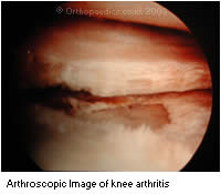 When the joint surface or articular cartilage has completely worn away to bare bone and the knee is painful, swollen and stiff it can be said that arthritis is present. Arthritis of the knee may affect 1, 2 or 3 of the major areas or compartments of the knee. These areas are under the kneecap (patello famoral arthritis), between the major bone ends on the inside of the knee (medial arthritis) and the same bones on the outside (lateral arthritis).
When pain and swelling and stiffness cannot be controlled, by conservative or non-operative measures such as limiting activity and the taking of medication, or when those limitations are severe, it is time to consider surgery. There are several different types of surgical techniques which are possible according to the exact nature of the problem.
Uni-compartmental Knee Replacement
More Information about Uni-compartmental Knee Replacement
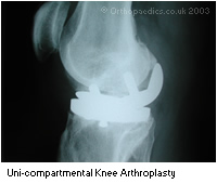 When the arthritis is severe in one compartment of the knee either the inner medial aspect or the lateral; outer aspect of the knee. Only a single side may be replaced in a uni-compartmental knee replacement. This may be particularly indicated in a young patient (under 65) so as to preserve as much of the patients original knee as possible. A uni-compartmental replacement of the knee can be performed by a minimally invasive technique where the incision is less than 10cm in length. This is often associated with a much quicker recovery for the patient with less pain stiffness, a shorter hospital stay and a more rapid return to activity, work, driving and to sport.
Patello-femoral Replacement
In situations where the arthritis is severe, it only affects the patello-femoral joint, the patient is young, incapacitated and the other techniques of patello-femoral realignment have failed then replacement of the patello-femoral part of the knee alone is possible. However the success of this particular technique is still as yet unproven, and it is only indicated in very specific cases.
Total Knee Replacement
More Information about Total Knee Replacement
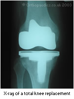 When the arthritis is severe, or affects more than one compartment of the knee. Where there is rheumatoid arthritis or in older patients, the whole knee joint is removed and replaced in a total knee replacement. In very special cases this may be necessary in younger patients when a special type of knee replacement will be used. New technology, new techniques and new types of knee replacements have made this procedure much more successful in recent years and the results are now as good or better than hip replacement .
Total knee replacement can now also be undertaken using minimally invasive techniques. The incision for knee replacement may now be less than 12cm in length. The surgery involves replacing the joint surfaces with a metal and plastic surface to all 3 compartments. The main ligaments are preserved and the leg straightened (if bowed) during the procedure. The range of movement is usually improved following surgery. The artificial joint usually bends from a straight position to at least 110 degrees. About 5-7 days are required in hospital. Pre-operative and post-operative physiotherapy is essential for success. The relief of arthritis pain is usually quickly apparent.
< BACK to Indications / Contra - indications | NEXT: Further Therapy >
Related Links..
+ How to make an appointment
+ Early Arthritis of the Knee - see all links
+ Back to Patient Information Home
+ See the clinic
+ More about Mr Johnson
+ top
© The Bristol Orthopaedics and Sports Injuries Clinic 2003. The Bristol Knee Clinic is a trading name of the Bristol Orthopaedic Clinic Ltd. privacy / copyright | contact | Powered By Create Medical